

This study uses agent-based modeling to examine how mechanical interactions between the extracellular matrix (ECM) and neurons shape neurite growth patterns and synapse formation. The ECM is the cellular environment around neurons and is crucial to brain development and function across life stages. The ECM is nowadays known to regulate axonal guidance, growth cone adhesion, synaptic plasticity, and more. Although many chemical and mechanosensing pathways have been identified, the mechanical components of these interactions remain difficult to isolate. Our underlying goal is to identify fundamental physical mechanisms underlying early brain tissue development, emphasizing environmental interactions. This research project is a part of the “Exploring Brain Mechanics” (EBM) CRC, which is dedicated to discovering the role of mechanics in the function and development of the brain.
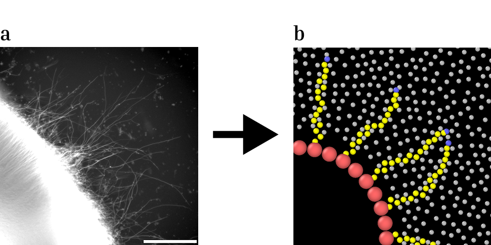
Our agent based approach models interactions with the environment by tension pulling of the growth cones, and deformation of the ECM by excluded volume interactions. This simplified approach both gives us a small set of parameters to adjust, and to tie these parameters to observations in experimental counterparts. In one such case, we collaborated with the groups of Marisa Karov and Sven Falk to compare our simulation model to experimentally observed growth patterns [1]. In this study, they investigated three gene mutations linked to neurodevelopmental disorders, using human brain organoids, growing on an artificial extracellular matrix (MatriGel). These mutations exhibited aberrant long-range connections, leading to premature loss of growth directionality compared to their wild-type counterparts. For the MID1 gene mutation, our model simulations revealed that increasing the growth cone adhesion angle was sufficient to replicate this altered growth pattern, suggesting that a malformed growth cone could be the cause of these alterations.
More recently, we collaborated with the group of Ben Fabry to investigate axonal growth patterns in three dimensional collagen networks, using hippocampal rat neurons [2]. From the experiments, we showed that in stiffer collagen axons tended to grow straighter, thus travelling greater distances in the same timeframe. Using our simulation model, we demonstrated that such growth patterns could result purely from passive interactions with the environment, thus reinforcing the importance of elementary physical forces in driving neuronal growth. Finally, we extend our model beyond the scope of elementary brain tissue formation, and test it with experiments of zebrafish spinal cord repair in collaboration with the groups of Silivia Budday and Daniel Wehner [3]. In these experiments, we observed axons as they grow through an induced wound lesion in the zebrafish spinal cord, in order to repair the damage. In such complex in vivo systems, additional mechanosensing effects were considered in order to enable us to recapitulate the growth patterns observed.
Some References
- Aberrant formation of long-range projections across different neurodevelopmental disorders converges on molecular and cellular nexuses Federica Furlanetto, Alejandro Segura, Mathar Kravikass, Pritha Dolai, Sarah Frank, Sören Turan, Angelica Luna Leal, Stephan Käseberg, Susann Schweiger, Chichung D. Lie, Vasily Zaburdaev, Marisa Karow, Sven Falk bioRxiv 2025.04.15.648981; doi: https://doi.org/10.1101/2025.04.15.648981
- In silico neuritogenesis model underpins mechanical interactions with extracellular matrix as determinants of persistent axonal growth in stiffer microenvironments Mathar Kravikass, Lars Bischof, Kristina Karandasheva, Federica Furlanetto, Pritha Dolai, Sven Falk, Marisa Karow, Katja Kobow, Ben Fabry, Vasily Zaburdaev bioRxiv 2026.03.13.708543; doi: https://doi.org/10.64898/2026.03.13.708543
- In silico model of axonal pathfinding during spinal cord regeneration in zebrafish larvae Oskar Neumann, Mathar Kravikass, Nora John, Rahul Gopalan Ramachandran, Paul Steinmann, Vasily Zaburdaev, Daniel Wehner, Silvia Budday bioRxiv 2026.04.17.719187; doi: https://doi.org/10.64898/2026.04.17.719187